


Transmission Electron Microscopy (TEM) can be used to investigate the morphology, structure, and chemical composition of fossils and (bio)minerals down to the nanoscale.
Below are shown, from left to right: optical image of unicellular fossil, dark-field scanning TEM image of the cross section along the black line, and nanoscale mapping of carbon and iron in the same zone.
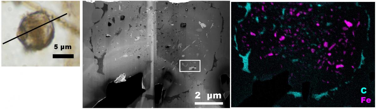
This has allowed us to demonstrate the presence of iron-bearing nanocrystals inside thick-walled unicellular microfossils and study the preservation of Paleoproterozoic microfossils (see publications). More generally, TEM allows to depict the nanoscale structure of fossils; it can find diagnostic morphological evidence (such as tri-laminar and/or finely ornamented walls) to distinguish fossil of small eukaryotic microorganisms from big fossil bacteria (see e.g. Javaux et al. 2004).
TEM is a highly versatile instrument performing several types of analyses.
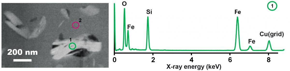
First, Energy Dispersive X-ray (EDX) spectroscopy can be performed on spots 1 to 10 nm in diameter, allowing identification and quantification of elemental composition at the nanoscale (below an example of EDX spectrum recorded in the green circle on the left, indicating an iron silicate).
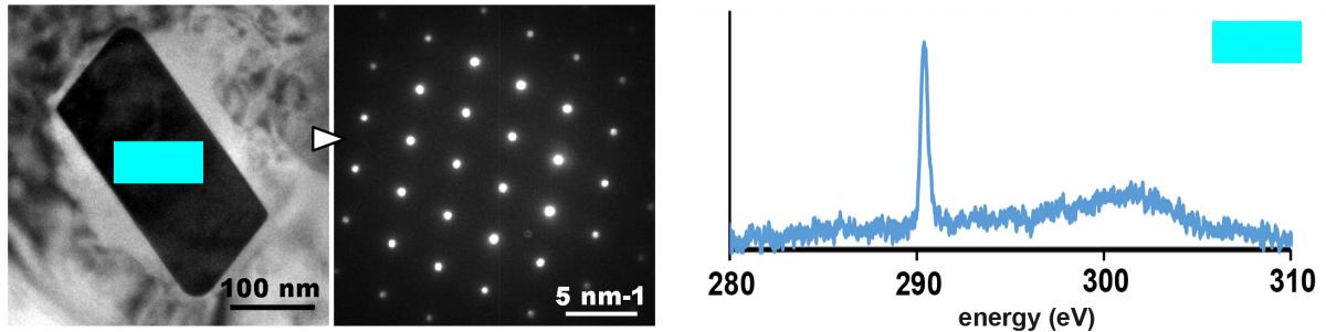
Second, TEM can perform diffraction at the nanoscale, allowing crystallographic identification of nanocrystals; an example of diffraction pattern identifying a siderite (Fe-carbonate) nanocrystal is shown below (left).
Third, TEM can characterize the speciation of elements, that is their molecular biding (C-C; C=C; C=O), their valence state (Fe2+, Fe3+), or their crystallographic environment (e.g. to distinguish silicon atoms in quarts against silicon in clays). This can be achieved using Electron Energy Loss Spectroscopy (EELS); an example of EELS spectrum identifying CO32- groups in the Fe-carbonate nanocrystal is shown below: this is very important to distinguish mineral carbon versus organic carbon at the nanoscale.
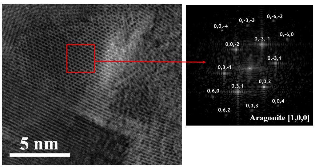
Fourth, TEM allows high resolution imaging of the atomic lattice columns (i.e. the distances between planes of atoms), as shown below (left); mathematical (Fourier) transforms of the high-resolution TEM images forms patterns of crystals large of e.g. 10 atoms (e.g. pattern on the right identifying aragonite nano-crystals). This allowed us to show microbially-mediated calcium carbonate nanocrystals in 2.7 billion years-old stromatolites (Lepot et al., Nature Geoscience 2008).
Our analytical platform is the Centre Commun de Microscopie de Lille