


Focused Ion Beam
Ultrathin sections (150 nm thick or less) are required to allow transmission of electrons (for Transmitted Electrons Microscopy) and X-rays (for Scanning Transmission X-ray Microscopy).
This technique uses beams of Gallium+ ions several nanometers large to make very localized craters in solid samples (rock, water ice, polymer,…). It allows the etching of patterns of any desired shape, including an ultrathin slice as shown in the schematic below. It is our preferred sample preparation method for Nanoscale (spectro)microscopy as it allows the sectioning of fossils together with their mineral matrix.

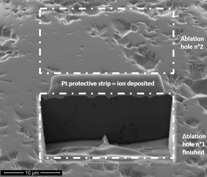
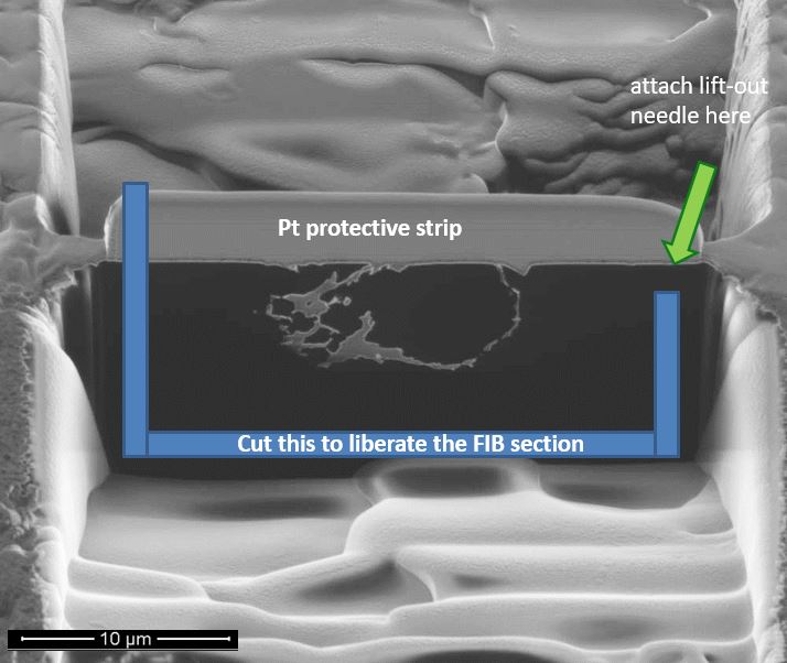
During the etching, the sample can be imaged with detection of the ions or electrons ejected (sputtered) in the process, or with secondary emission / or backscattering of electrons beams (like in a Scanning Electron Microscope), as shown in the images below. The image on the right shows a FIB-etched rectangular crated. The second image shows that with a second rectangular crater, a cross-section can be produced. This cross section can then be liberated from the sample by etching the section along the blue lines. Then, we stick it onto a thin needle with a platinum spray, transport the section in contact with copper grid, and stick in again with platinum on the grid. The needle is then liberated by FIB-etching of its platinum bond. The FIB section is finally polished with low intensity, grazing ion beams. The full FIB preparation process is shown in this video by Tomáš Hrnčíř.
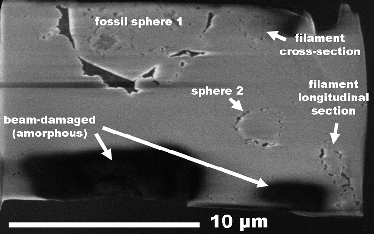
Below is shown an example of a typical FIB ultrathin section cut across 4 microfossils (organic matter appears in black) embedded in a quartz (grey) matrix. The typical area of the regions thinner than 100 nm is usually 15x10 micrometers. In quartz and many other minerals, larger areas tend to bend due to mechanical constraints, which leads to irregular etching forming much thinner areas (as shown in the dark zones at the bottom on this FIB-section). These thinner areas are usually amorphous.
We use the Frech RENATECH network analytical platform at Lille's IEMN.