


Scanning Electron Microscopy (SEM) uses a beam of electrons to image the topography of the surface of samples (secondary electrons), to image chemical contrast (backscattered electrons), and to perform elemental quantification and mapping using Energy Dispersive X-ray Spectroscopy (EDXS).
SEM imaging has a sub-micrometric lateral resolution, down to ~20 nm on our instrument. EDXS elemental analyses/maps have a 500 nm to 1 µm lateral resolution depending on electron acceleration voltage.
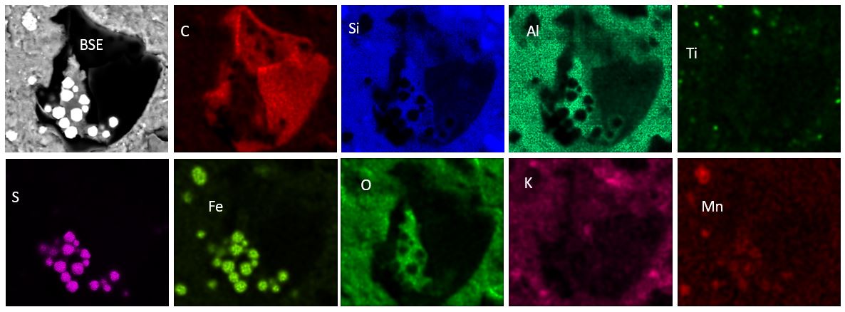
This technique is crucial to analyse the chemical composition of sedimentary rocks. Below is shown an example of a microfossil cropping out at the surface of a thin section cut parallel the bedding plane of a mudstone. In the Backscattered electrons image (BSE), the low atomic number zones (e.g. carbon-rich microfossil) appear darker than the high atomic number zones (e.g. Fe- or Ti-rich), as confirmed with the elemental mappings. This microfossil was trapped between fine-grained layers of Al-silicates with abundant TiO2 nanograins, and associated with pyrite (FeS2) that has been partly oxidized to Fe-Mn-oxides.
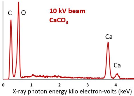
X-ray (EDXS) spectra allow the quantification of elements. In the spectrum below, recorded on calcium carbonate, the surface of each band is proportional to the abundance of this element in the sample. The detection limit is about 0.5-1 %.
We use a FEI Quanta 200 SEM equipped with a Bruker Quantax EDXS available at LOG. This equipment operates in low vacuum to analyse materials without conductive coating.
Contact : Philippe Recourt.